Introduction to the main light source in the gel imaging system, the gel imaging system, as the name implies, is an optical system that can image the gel, and the light source is an important part of its optical path system. It can be said that there is no light source, and the imaging system is almost Can not be used, but compared with CCD, lens and analysis software, often did not get the attention it deserves. We often encounter users who know that there is a 305nm and 365nm UV lamp in the gel imaging transmission when the system light source fails or the entire system is scrapped, or it is unclear what is the difference between them. Of course, there are sellers who are not in place after sale, but it also reflects our lack of attention to the light source of the gel imaging system and lack of sufficient understanding. We have written this article to help users clarify their differences and their respective roles. , giving you a better choice of buying, using and maintaining a gel imaging system.
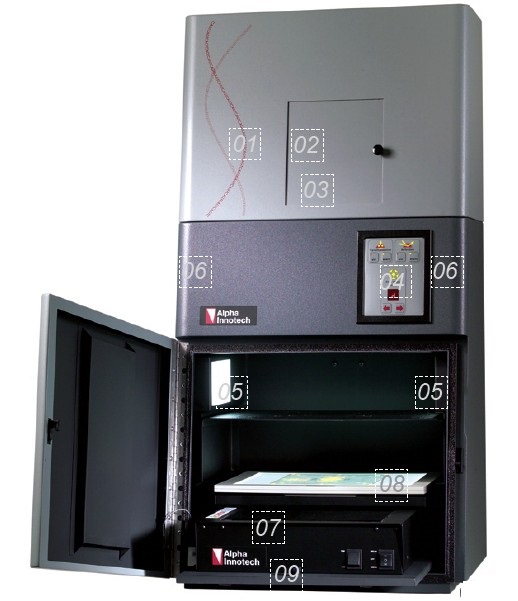
Depending on the application, different types of gel imaging products are often equipped with different light sources. The general types can be divided into white light source, ultraviolet light source and high-intensity three-color LED light, in which white light and ultraviolet light source respectively have transilluminate and Reflection (Epi). As shown in the figure below, we will introduce the position and characteristics of each light source by taking the DE-500 multi-function black box with the well-equipped ALPHA INNOTECH Fluorchem HD 2 as an example.

1, stainless steel material DE-500 dark box body; 2. The location of the CCD; 3. The location of the lens; 4, the body light source control panel, can control the filter wheel position and the excitation light source switch; 5. Double-sided reflective white light source; 6. Double-sided dual-wavelength (254nm, 365nm) reflective ultraviolet light source; 7. Height-adjustable pull-out tray for chemical lighting use; 8, transmission white light board, foldable; 9. Dual wavelength (302 nm, 365 nm) ultraviolet transmission source; In addition: In the Fluorchem Q multi-color fluorescent line system, three-color LED light source is also installed on both sides. |
Below we briefly introduce the various light sources and their uses of the gel imaging system. The first is the double-sided reflective white light source, which is also the most used light source. It is usually used as a system illumination source , that is, to place or adjust the position of DNA glue or protein glue. Or use it when adjusting the focus of the lens. Of course, it is necessary to turn on the reflected white light when imaging non-transparent material gelatin or other imaging substances.
Secondly, it transmits white light, which is commonly used as a white light board. It is mainly used for protein gel shooting . Because the white light board is located above the transmissive UV light box, it does not affect the use of the transmitted ultraviolet light source, and also saves indoor space. The white light board can generally Folded upright at the back of the cabinet.
Transmitted ultraviolet light source, ie UV transmissive light box, is mainly used for DNA gel shooting . Since the fluorescent dye commonly used in DNA is EB (ie Ethidium bromide, ethidium bromide), it is excited by a standard 302nm UV transilluminator and emits orange-red signal. It can be photographed by a gel imaging system with a CCD imaging head, so the wavelength of the transmitted ultraviolet light is usually 305 nm.
At present, many manufacturers have designed dual-wavelength transmissive UV lamps (302nm, 365nm). For example, the famous American Alpha INNOTECH company (see ), its 365nm light source is mainly suitable for preparation observation and cutting strips. Reduce damage to DNA and reduce damage to people.
The source of the UV-reflecting lamp is generally an optional accessory. The main reason is that it is not widely used, and it is only required to be imaged in a non-transparent material such as a DNA carrier such as paper tomography. Often used are transparent agarose gel and polyacrylamide gel, the transmission source can be fully satisfied, so most manufacturers only use the UV-reflecting light source as an option, but the position and interface are reserved on the instrument.
It should also be pointed out that the use of pull-out trays, because chemiluminescence imaging does not require any light source, is self-luminous imaging, but because the chemiluminescence signal is relatively weak and unstable, the lens of high-grade chemiluminescent gel imaging system is often Manual focus, so a height-adjustable tray is required for chemiluminescence imaging.
In addition, the three-color LED light source mentioned above is three high-intensity reflection exciters (475 nm, 534 nm, 632 nm), which is an indispensable component of multi-color fluorescence imaging systems. Fluorchem HD 2 's DE-500 black box also reserves a continuous wavelength source fiber interface for future expansion to continuous wavelength sources (excitation wavelength range of 400-750nm), covering some fluorescence that requires special excitation light, mostly used in many Color fluorescence (Cy3/Cy5) and in vivo imaging.
Morel Mushrooms,Fresh Morels,Freezing Morel Mushrooms,Morels Edible Fungi
Guangyun Agricultural Biotechnology (Jiangsu) Co., Ltd , https://www.7-mushrooms.com